
Biosensors are exclusively of analytical nature, designed to measure in a quantitative fashion diverse physicochemical parameter. In industrial and environmental settings, biosensors find applications in evaluating air, soil and water quality, among others. For biological systems in research and medical settings, current biosensor designs involve sensing elements to measuring metabolic products, pH, cell adhesion forces, specific pathogen biomarkers, among others. The key design elements are high target molecule specificity and sensitivity being superior than standard assays provide. Particularly in the interest of health care, biosensor platforms able to detect ultra-low concentrations of diverse disease biomarkers facilitate early disease detection, rendering timely treatments possible. Due to the inherent label-free and rapid operation, biosensors are increasingly applied in point-of-care diagnostics.
RESEARCH & DEVELOPMENT PROJECTS
⦿ nanowire Biosensor for Ultra-sensitive and highly selective Disease Biomarker Detection

Field-effect transistors (FET) based biosensors are vital in life science applications, as they offer direct, fast, and highly sensitive label-free detection of diverse biomarkers of interest. In this work, we investigate different surface functionalization approaches and the covalent immobilization of biomarkers using biocompatible ethanolamine and poly-(ethylene glycol) derivate coatings, in order to substantially increase both the sensitivity and molecular selectivity of nanowire-based FET biosensor platforms. Significantly enhanced binding specificity, biomarker density, and target biomolecule capture efficiency were achieved for ssDNA as well as for proteins for human pathogens. This methodology was applied to InP nanowires that due to their low surface recombination rates were used as new active transducers for biosensors. The developed devices provide ultrahigh label-free detection sensitivities ∼1 fM for specific ssDNA sequences, measured via the net change in device electrical resistance. Similar levels of ultrasensitive detection of ∼6 fM were achieved for a Chagas Disease protein marker (IBMP8-1). The developed InP nanowire biosensor provides thus a qualified tool for detection of the chronic infection stage of this disease, leading to improved diagnosis and control of spread. These methodological developments are expected to substantially enhance the chemical robustness, diagnostic reliability, detection sensitivity, and biomarker selectivity for current and future biosensing devices. >Download Study<
⦿ Highly-sensitive and label-free indium phosphide thin film FET biosensor

The development of highly-sensitive and label-free semiconductor-based, biomarker-detecting sensors has important applications in areas such as environmental science, biomedical research and medical diagnostics. In the present study, we developed an Indium Phosphide (InP) semiconductor-based resistive biosensor using the change of its electronic properties upon biomaterial adsorption as sensing element. To detect biomaterial at low concentrations, the procedure of functionalization and covalent biomolecule immobilization was also optimized to guarantee high molecule density and high reproducibility which are prerequisite for reliable results. The characterization, such as biomolecular conjugation efficiency, detection concentration limits, receptor:ligand specificity and concentration detection range was analyzed by using three different biological systems: i) synthetic dsDNA and two phytopathogenic diseases ii) the severe CB-form of Citrus Tristeza Virus (CTV) and iii) Xylella fastidiosa, both causing great economic loss worldwide. The experimental results show a sensitivity of 1 pM for specific ssDNA detection and about 2 nM for the specific detection of surface proteins of CTV and X. fastidiosa phytopathogens. A brief comparison with other semiconductor based biosensors and other methodological approaches is discussed and confirms the high sensitivity and reproducibility of our InP based biosensor which could be suitable for reliable early infection diagnosis in environmental and life sciences. >Download Study<
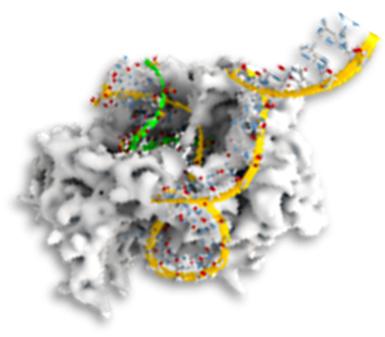
⦿ cell force sensors reveal adhesin holdfast of single bacteria and biofilm communities

Single cell adhesion to a surface is the first and critical step to form a biofilm. Specific extracellular components facilitate adhesion by reducing the energy barrier formed between approaching surfaces, in a process which depends on bacteria-host adaptation mechanisms. It is thus important to probe the specific dependence of bacterial cell adhesion on surface chemical composition and the corresponding molecular mechanisms. Further complexity to the scenario of bacterial biofilm infection arises from the ability of many species to modulate cell adhesiveness by specific transmembrane adhesion proteins, and hence host colonization, in response to changing environmental conditions. We employed spatially ordered nanowire arrays to evaluate Xylella fastidiosa single cell adhesion forces and explore their dependence on chemical surface compositions. Modulation in cell holdfast in dependence of a transmembrane adhesin, XadA1-functionalized nanowire can arrays directly assess the impact of XadA1 on cell-surface interaction (Figure), mimicking its presence in outer membrane vesicles (OMV), which are released in living environment. The adhesin significantly increased the cell holdfast forces up to ~45 nN, suggesting that the protein is a promoter of cell adhesion. From the biological point of view, adhesins provide important implications in biofilm fitness and pathogenicity. >Download Study<
⦿ Nanowires as Higly-sensitive cell Force Sensors with Super-resolution Position Detection

Many techniques have been developed for measuring cell adhesion forces. Commonly, flow chamber and AFM techniques are used. However, single point force measurements cannot provide a broader picture of high data content of force networks of whole cells or multicellular tissues. Recent studies have demonstrated the use of force sensors based on vertical nanopillars for mammalian cell studies, and nanopillar deflections imposed by cells were commonly measured by electron microscopy in dry samples, with possible tension effects. Here, we developed indium phosphide (InP) nanowire arrays to the measure accurately bacterial adhesion forces which exhibit forces over magnitude lower than for mammalian cells. The deflection of nanowires was obtained with localization subdiffraction technique in confocal laser scanning microscopy measuring the reflected laser intensity at the nanowire apexes to track their position over time. Adhesion forces of the phytobacteria Xylella fastidiosa were then calculated from the deflection data using linear elasticity theory. The center of the nanowire intensity profile was calculated using 2D Gaussian fitting; the uncertainty of was estimated, under consideration of the thermal limit, as low as 6 nm using a localization microscopy concept. We demonstrate that this methodology can measure bacterial adhesion forces in situ with a lower limit of ~5 nN. >Download Study<
⦿ Single-Crystal TiO2 force Transducers allow monitoring of high-speed rotation dynamics

Optical trapping of (sub)micron-sized particles is broadly employed in single-molecule methodologies. The materials commonly employed for these particles, however, have physical properties that limit the transfer of linear or angular momentum (or both). This reduces the magnitude of forces and torques, as well as the spatiotemporal resolution, achievable in linear and angular optical traps. Here, we overcome these limitations by the use of single-crystal rutile TiO2, a material with an exceptionally large optical birefringence, a high index of refraction, good chemical stability, and is amenable to geometric control at the nanoscale. We show that rutile TiO2 nanocylinders form powerful joint force and torque transducers in aqueous environments that achieve nN·nm torques at kHz rotational frequencies at nanometer dimensions. We demonstrate that rutile TiO2 nanocylinders outperform other materials and offer unprecedented opportunities to expand the control of optical force and torque. We complement our fabrication approach with a functionalization strategy that achieves dense, uniform, and area-selective coating to couple a variety of biomolecules while providing particle monidispersity >Download Study<
⦿ Development of single probe DNA molecule surface patterns with nanometer accuracy

The ultimate physical limit in building organic and biological nanostructures can be reached by assembling one by one molecule in a precisely controlled manner. Previously established concepts rely on an irreversible biomolecule delivery process, but provides no proofreading of the delivery. In this work, we demonstrate the directed delivery of single DNA molecules via an Atomic Force Fluorescence Microscope using a procedure that intrinsically allows a reversible positioning by using specific molecular interactions between complementary DNA oligonucleotides. This methodology allows for a simple “drag-and-drop” procedure, which relies on the statistical breakage of the molecular interactions under a certain force load and does not require any external optical or electrical triggering. The developed method can further be used for other (bio)molecules and nanoparticles by modification of the respective transport unit. In addition, deposited molecules can serve as template for further self-assembly of other components using specific molecular recognition. >Download Study<