
HEMINE CHLORIDE CRYSTALS
Color enhanced SEM image of a “cracked riverbed” with differently grown hemine crystal structures. The Nobel prize winning (1930) iron-containing porphyrin molecule is the essential part of Animalia respiratory gas transport system and serves often as “x-factor” for bacterial growth. The shown image of a dried aqueous hemin chloride solution exhibits different coexistent asymmetric crystalline growth shapes.
Material: Hemine chloride on silicon wafer
Preparation: 1:1 Hemine chloride:H2O on Si 12 h at RT. Dried by evaporation.
Measurement: SEM imging using Phenom ProX, 1540x magnification.
Finalist in MRS Science as Art Awards 2013
Credits: Richard Janissen
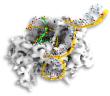
TREFOIL DNA-PROTEIN COMPLEX
For gene therapy and DNA vaccination, recombinant proteins are designed for plasmid DNA delivery to the centrosome of mammalian cells. For that purpose, artificial DNA binding sequences were fused to recombinant human dynein light chain domain LD4. The AFM topography reflects an eukaryotic pVAX1 plasmid-DNA (green) starting to complex at highly negative charged sequence regions with the positive charged dynein light chain domain LD4 (brown), forming compact particles of ~ 100 nm at higher concentrations.
Measurement: Acoustic AC-mode (AAC) imaging on dried sample on mica using an Agilent AFM5500. 600×600 nm scan, 6 nm full Z-scale.
PicoCafe AFM calendar contest winner 2014
Credits: Richard Janissen


NANOWIRE STARS IN THE NIGHT SKY
One-dimensional semiconductor nanostructures are considered in many applications. In biophysics, such nanowires represent also excellent biosensors as the change in local charge density of the FET upon target molecule binding allows detection sensitivities down to femtomolar concentrations. A common technique for growth poses the vapor-liquid-solid (VLS) method where metal colloids (such as gold) are used as catalysts. In the shown color-enhanced SEM image, the colloids undesirably aggregated prior the VLS growth leading to unserviceable, but beautiful star-like structures.
Material: Indium Phosphide
Measurement: Field Emission Scanning Electron Microscopy – FEI Inspect F50.
Credits: Douglas S. Oliveira & Richard Janissen
SENSING CELL FORCES VIA NANOWIRES
The image shows a color-enhanced SEM image of a phytopathogenic bacteria attached to an Indium Phosphide nanowire array. Surface attachment of a planktonic bacteria, mediated by transmembral adhesins and extracellular polymeric substances (EPS), is a crucial step for biofilm formation, impacting host colonization and virulence. Such nanowire arrays are used to dissect factors involved in the early stage biofilm formation of the phytopathogen Xylella fastidiosa. Ex-vivo force sensor experiments demonstrate single-cell adhesion forces up to ~50 nN, rendered by the bacterial adhesins and EPS secreted matrix.
Measurement: Field Emission Scanning Electron Microscopy – FEI Inspect F50.
Figure1a International Science Art Contest Finalist, Exhibition in Switzerland 2022
Credits: Prasana Sahoo & Richard Janissen

PROKARYOTIC RNA POLYMERASE
The image depicts the authors imagination of E. coli DNA-dependent RNA polymerase during transcription. In all living organisms, this Nobel Prize (1959) winning protein complex regulates the production of necessary proteins involved in cell metabolism, cell division, genome repair, and immunological response. Therefore, as this enzyme is highly structurally and functionally conserved in all three domains of life, RNA polymerases constitute a primary pharmaceutical target for many pathogenic bacteria and viruses. The RNA chain elongation is stochastic and highly dynamic, interrupted by pauses that represent a gene regulatory feature: they facilitate the timely interaction of regulatory factors and the synchronization with the subsequent translation. Despite many studies, the current mechanistic and kinetic model of transcriptional pausing remains controversial.
Credits: Richard Janissen
RNA VIRUS EV-A71
The image shows the authors interpretation of a RNA virus of the family of enteroviruses during RNA genome replication. Enterovirus is a genus of positive-sense single-stranded RNA viruses associated with several human and mammalian diseases. Serologic studies have distinguished 71 human enterovirus serotypes. Enteroviruses cause a number of infectious illnesses which are usually mild. The most common enteroviruses are echovirus, coxsackievirus, and poliovirus. Most illnesses caused by enteroviruses are mild but more severe diseases can sometimes develop in certain patients, including the central nervous system and heart conditions, pneumonia and hepatitis. Commonly, these viruses can spread to other organs such as the spleen, liver, bone marrow, skin and heart.
Credits: Richard Janissen


DEADLY 'TREE' OF PHYTOPATHOGEN XYLELLA FASTIDIOSA
The color-enhanced fluorescence image shows a fractal-like arrangement of the bacteria Xylella fastidiosa, adhered on Indium Phosphide. X. fastidiosa is a vascular phytopathogen causing diseases worldwide in important crops (e.g., coffee, grape, citrus, among others). The bacteria colonizes two distinct habitats: the xylem vessels of plants and the foregut of xylem-feeding sharpshooters, which are transmission vectors. This species shares genetic traits with other human bacterial pathogens and has slow duplication time of ~6 h, which renders it a good model for studying the stepwise biofilm formation and encapsulation in EPS. As many other gram-negative bacteria, this species uses twitching motility to colonize surfaces that plays a crucial role in biofilm development.
Material: Xylella fastidiosa pauca strain, Indium Phosphide substrate
Preparation: 24 hours growth of bacteria in Periwinkle Wilt media
Measurement: Leitz Ergolux upright confocal microscope, 20x
Credits: Silambarasan Anbumani (UNICAMP)
FRACTAL PATTERN OF MIN PROTEIN REACTION-DIFFUSION SYSTEM
The animation exemplifies the reaction-diffusion system of Min proteins. In E. coli bacteria, Min proteins make sure the cell divides evenly in two halves. In an in vitro setting, they have the capacity to form a variety of beautiful patterns, as demonstrated by this spiral.
Method: MinE protein on a supported lipid bilayer (MinD and ATP added).
Measurement: Spinning disk confocal microscopy
Credits: Sabrina Meindlhumer (TU Delft)
