
Chemo/Biointerface science studies and controls molecular interactions at surfaces and in polymers. The motivation for chemo/biointerfaces stems from the urgent need to increase the understanding of interactions between molecules and (in)organic materials. Nowadays, chemo/biointerfaces are a critical component of diverse revolutionary applications in biotechnology, clinical diagnostics, tissue engineering, novel drug carriers, novel composites, and fundamental research. Developments in chemo/biointerfaces are inspired by nature’s principles, but aim at creating novel hybrid materials with tailored functionalities, in order to understand, predict, and design physicochemical interactions at the interface with biomolecular and polymer systems. The rich diversity of function provided by natural or synthetic chemo/biomolecules at interfaces opens unparalleled opportunities for novel applications in life science, health care and chemo/biocomposites.
RESEARCH & DEVELOPMENT PROJECTS
⦿ mechanophores can reveal quantitative information of internal stresses in polymers

Mechanophores are powerful molecular tools used to track bond rupture and characterize mechanical damage in polymers. The majority of mechanophores are known to respond to external stresses, and we report in this study the first precedent of a mechanochemical response to internal, residual stresses that accumulate during polymer vitrification. While internal stress is intrinsic to polymers that can form solids, we demonstrate that it can dramatically affect the mechanochemistry of spiropyran probes and alter their intramolecular isomerization barriers by up to 70 kJ mol–1. This new behavior of spiropyrans (SPs) enables their application for analysis of internal stresses distribution and their mechanochemical characterization on the molecular level. Spectroscopy and imaging based on SP mechanochemistry showed high topological sensitivity and allowed us to discern different levels of internal stress impacting various locations along the polymer chain. The nature of the developed technique allows for wide-field imaging of stress heterogeneities in polymer samples of irregular shapes and dimensions, making it feasible to directly observe molecular-level manifestations of mechanical stresses that accompany the formation of a vast number of solid polymers. >Download Study<
⦿ Homogeneous & Biocompatible Coating of Graphene Oxide for Biomedical Applications

Due to its unique material properties, graphene oxide (GO) has immense potential for diverse biomedical applications, including organoid bioprinting, drug delivery, nanocomposites, and biosensors. However, implementation of GO in new applications is being prevented due to an inability to coat this material appropriately. Functional coating of GO requires high molecule density, coating homogeneity, chemical stability, and biocompatibility and current methodologies do not fulfill these requirements. We have solved the current limitations by developing a powerful wet-chemistry functionalization method. The procedure employs two monolayer coatings covalently attached to the entire GO lattice. The first monolayer consists of colamine, which couples via ether formation to the abundant hydroxyl groups on the GO surface. The second monolayer is realized by covalently binding biocompatible, heterobifunctional poly(ethylene glycol) to colamine, which can be further exploited to covalently immobilize pharmaceutical or biological molecules. With a step-wise characterization of the process, we show that this method paves the way for GO to be used in future in vivo biomedical applications. >Download Study<
⦿ covalent DNA anchoring for enhanced single-molecule temporal and force stability

Single-molecule techniques have become a valuable tool in real-time quantitative investigation of biomolecular processes under applied force. However, high forces may be required, e.g. to unfold proteins, or to probe the force range of proteins that actively transcribe or package the genome. Frequently, the application of high forces decreases the sample lifetime, hindering data acquisition. To provide experimentally viable sample lifetimes in the face of high pulling forces, we designed a novel DNA-tethering strategy. Our approach exploits covalent functionalization based on heterobifunctional poly(ethylene glycol) crosslinkers, allowing to strongly tether DNA while simultaneously suppressing undesirable non-specific adhesion. Compared to more commonly employed anchoring strategies, this method allows 3-fold higher pulling forces (up to 150 pN) and exhibit up to 200-fold higher lifetimes (exceeding 24 h at a constant force of 150 pN). This advance makes it possible to apply the full range of biologically relevant force scales to biomolecular processes, and its straightforward implementation should extend its reach to amultitude of applications in the field of single-molecule force spectroscopy. >Download Study<
⦿ covalent biomolecular coating of single-crystal titanium dioxide nanocylinders for OT

Single-crystalline titanium dioxide (TiO2) is a new material finding its application in single-molecule optical (torque) tweezers due to its exceptionally large optical birefringence, which makes it an excellent candidate for incorporation into torque transducers. For full implementation of TiO2 as force and torque transducer in single-molecule techniques, it is key to achieve chemically robust and highly densed surface functionalization and bioconjugation. However, surface functionalization of TiO2, using common alkoxysilane surface linkers, is known to be less efficient in terms of linker density and uniformity than other oxide materials (e.g. SiO2, Al2O3). Here, we present a successful top-down fabrication and surface functionalization of single-crystal TiO2 nanocylinders with diameters in the range of 100-200 nm. The applied surface coating, consisting of two monolayers of mono-reactive surface linkers and biocompatible poly(ethylene glycol), allow stable DNA attachment while providing nanoparticle monodispersity in physiological solutions. We demonstrate the use of these coated TiO2 nanocylinders by stretching and twisting individually tethered DNA molecules. >Download Study<
⦿ functionalized Titanium implants with cyclic RGD-peptide for bone marrow integration

The fixation of cementless endoprostheses requires excellent fixation at the bone implant interface. Although the surface structures of these implants are designed to promote osteoblastic differentiation, poor bone quality may prevent or delay osseointegration. There is evidence that cyclic RGD peptides known as recognition motifs for various integrins, promote cellular adhesion, influence cellular proliferation, and differentiation of local cells. However, previous methods used non-covalent strategies to attach cRGD peptides to biometals and did not consider to increase the metal surface biocompatibility in respect to non-specific cell adhesion. Here, we functionalized titanium, cobalt chrome, and stainless steel with different surface topographies frequently used in hip arthroplasty with biocompatible poly(ethylene glycol) and cRGD peptides in a chemically stable, covalent fashion. We used an in vitro human bone marrow cell model to evaluate the osteogenic potential with respect to their proliferation and differentiation. In contrast to previous beliefs, our results demonstrate that cRGD provides no significant improvement in cellular proliferation and osteoblastic differentiation. >Download Study<
⦿ non morphology and function-affecting covalent immobilization of living bacteria

Preserving the morphology and (intra)cellular integrity is important during immobilization of living bacteria. The key reasons of this immobilization approach is to inhibit cellular autolysis, to preserve cellular components and morphology, and to present cells with a distinct microscopical appearance. Current methods use additive or denaturing fixation solutions, while both groups lead to significant changes in the native morphology, biomolecule integrity, and cell death. Here, living phytobacteria cells were covalently immobilized to amine- or carboxyl-terminated functionalized glass surfaces and with non-specific adhesion-suppressing poly(ethylene glycol) to couple to cell outer- or transmembrane proteins and glycoproteins. To verify that proteins are still in their native conformation and localization, we successfully detected via fluorescence-labeled primary antibodies the localization of the TolB-Pal complex residing in the peptidoglycan layer, and the protein frnE, a disulfide isomerase chaperone, present throughout the cytoplasm. As this methodology provides the immobilization of living cells, single-molecule live cell imaging of motile and spinning bacteria will be feasible at last.
⦿ biocompatible, covalent surface coupling of biomolecules for snanning probe microscopy

Due to the exceptional sensitivity of single molecule detection and its ability to detect unique events in an ensemble of molecules it presents a huge potential for the study of biorecognition processes. However, due to problems associated with the sensitivity, reproducibility, long-term stability, and non-specific adhesion of biomolecules, biomolecule-functionalized surfaces are not widely available from commercial sources and are time consuming due to the complex procedures containing many pitfalls. In order to circumvent these problems we developed an optimized wet-chemistry procedure resulting in a high biomolecule density, homogeneous coverage, and a low unspecific background when binding nucleic acids, peptides, and proteins alike. We investigate and compare to existing protocols the bio-functionalization efficiency of different material treatments and chemical coupling strategies to silicon, silicon oxide, and silicon nitrite surfaces and AFM cantilevers. In comparison, our ubiquitous applicable, simple and reproducible functionalization procedure of biomolecules (DNA, peptides, and proteins) to oxidized surfaces yield an increase up to 3-fold in biomolecule binding efficiency. >Download Study<
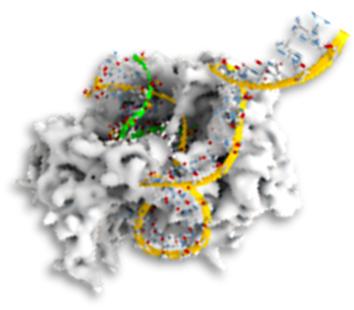
⦿ DNA curtains on gold nanopattern with lipid bilayer for unspecific adhesion suppression

In diverse single-molecule assays it is desirable to achieve both an ordered anchorage of biomolecules and a nonspecific adhesion suppressing surface. Here, we developed a methodology to bind specifically and in a covalent fashion DNA to gold nanostructures and create a surrounding lipid bilayer surface to prevent unspecific biomolecule adhesion. Inspired by previous pioneering work on DNA curtains, we focused here on the development of a Stretched Oriented DNA Arrays (SODA) microfluidic platform for visualization and characterization of protein-DNA and protein-protein interactions on a DNA strand at single-molecule level. Single DNA molecules are arranged in a pre-defined manner, stretched and bound to functional gold structures at the glass surface with a surrounding DOPC phospholipid bilayer. This configuration enables simultaneous visualization of the dynamics of labelled proteins on hundreds of DNA strands using TIRF microscopy. The platforms allows to e.g. monitor 1-dimensional motions of genome-processing machines, the effect of roadblocks and crowding on polymerases, the assembly kinetics of multi-protein complexes, mechanism of transcription-coupled translation, among other.
⦿ Lithography-based cell traps for live cell imaging in physiological environments

Controlling the adhesion landscape of living cells on a surface is emerging as a new tool for investigating fundamental mechanisms, such as differentiation, division, motility, and formation of bacterial biofilms. More excitingly, with the ability to capture and separate individual cell, single-molecule live cell imaging of intracellular processes would become more feasible. The key requirement of all these studies is the development of micro-patterns of various shapes and dimensions, and specific chemical surface modifications to either repel or attract cell in a deterministic way. Here, we used low-cost laser lithography to produce microscale patterns and different chemical coatings to confine bacterial cells within the pattern. We further immobilized covalently different (bio)molecules that induce cell binding and improves the hold-fast of the cell. Implemented in fluidic chambers, we demonstrate that this methodology can be exploited to e.g. investigate steps in bacterial biofilm formation and the stability of cell adhesion at real time via fluorescence microscopy. The cost and time effective production and easy scalability of these cell traps provide an alternative to the more complex microcontact printing methodology.